Personalized plastic surgery, laser & anti-aging care in Haeundae, Busan
en-US

Eye Shape, Fat Volume and Skin Thickness in Under-Eye Care
Why choosing the right under-eye procedure depends on lower-eyelid anatomy: thin periorbital skin, fat position versus volume, tissue elasticity and age.
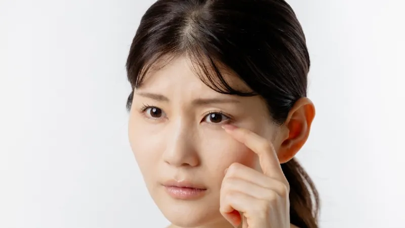
Eye Shape, Fat Volume and Skin Thickness in Under-Eye Care
Medically reviewed by Dr. Kim Ki-Tae, Board-Certified Specialist in Plastic Surgery, issued by the Korean Society of Plastic and Reconstructive Surgeons
Two people can walk into a clinic with the same sentence — "I look tired even when I'm not" — and leave with entirely different plans. That is not inconsistency. Choosing the right under-eye procedure is a question about structure, and the structure producing a shadow in one face is often not the structure producing an almost identical shadow in another. This article is about the factors an examination is weighing, and why each of them changes the answer rather than merely decorating it.
TAE Plastic Surgery Clinic in Haeundae, Busan runs three separate under-eye services within its eye plastic surgery category, and its stated position is that which one applies follows from assessment rather than from what a patient asks for by name. What follows explains the reasoning behind that position. It does not tell you which service fits you; no article can do that.
The Lower Eyelid Is Not Like the Rest of Your Face
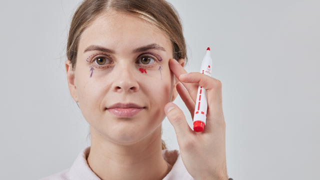
Every decision in this region starts from one anatomical fact. The clinic states that the periorbital area — the zone surrounding the eye — has thin skin with a dense concentration of blood vessels and nerves, and that this is precisely why eye shape, the quantity of fat and the thickness of the skin have to be considered together rather than one at a time.
Thin, richly supplied tissue behaves differently from the skin of a cheek or a jawline in three ways that matter.
It shows what is underneath it. A vessel, a shadow, a contour or a slight excess of fluid is visible through eyelid skin in a way it would never be elsewhere. This is why small structural differences produce disproportionately large changes in how rested a face looks.
It bruises and swells readily. Dense vasculature in a thin, loose envelope means that fluid and blood have somewhere easy to go. Early swelling and discoloration after anything done in this region are ordinary, not a sign of technical difficulty.
It leaves very little room for a mismatched plan. There is no thickness of tissue to absorb a slightly-wrong amount of anything. Removing a little too much or adding a little too much in a region measured in millimeters registers on the surface.
The clinical literature supports how exacting this area is. StatPearls, a peer-reviewed reference hosted by the National Library of Medicine, sets out in its chapter on transconjunctival blepharoplasty the lower eyelid in layers — skin and muscle in front, the orbital septum in the middle, retractors and conjunctiva behind — held in position by canthal tendons at each corner, and notes that the tarsal plate itself measures around a millimeter in thickness. It states plainly that the procedure depends on the surgeon's abilities as an examiner and diagnostician, not only as an operator.
Fat Volume and Fat Position Are Separate Findings
"Fat" is treated in casual conversation as one variable — too much or too little. In the lower eyelid it is at least two, and they are not the same problem.
How much fat is present is a volume question. Volume can be reduced, distributed unevenly between the compartments, or diminished across the whole area, which reads as hollowness rather than as bulging.
Where the fat sits is a position question. The same quantity of orbital fat can sit contained behind the septum or press forward past the orbital rim, producing a visible ridge with a groove beneath it. A groove of that kind is often not an absence of tissue at all; it is the boundary where a forward bulge stops.
The reason this distinction earns its own section is that a person can present with both at once — a fullness above and a hollow below — and the hollow can be an illusion cast by the fullness, or a genuine loss of volume, or both together. Those are different findings with different implications, and no amount of confidence about which one you have replaces looking.
The StatPearls account is useful here too, because it sets out how much anatomical detail sits behind a simple-looking bulge. The lower eyelid has medial, central and lateral fat pads; the inferior oblique muscle runs between the medial and central pads; and a thickening in the septum called the arcuate expansion separates the central from the lateral pad. It also records that the lateral pad is the one most often under-addressed by inexperienced surgeons. Those are not patient-facing details, but they explain why the assessment is regional and specific rather than a single judgment about "bags."
Skin Thickness and Elasticity Decide What the Tissue Does Afterward
If fat explains the shape, skin explains what happens to that shape once anything is altered.
Skin has to redrape. When volume beneath it is reduced, added or moved, the surface must accommodate the new contour. Skin with good elasticity contracts and settles onto it. Skin that has lost elasticity does not reliably do so, and a technique that suits the first situation may not suit the second at all.
Skin thickness also changes what shows. Thinner skin transmits color and contour more directly, which is why some darkness under the eye is not pigment or shadow but the visibility of what lies beneath. Thicker skin conceals more and settles differently.
Both properties change with time. MedlinePlus, run for patients by the National Library of Medicine, records that skin becomes thinner and loses fat as we age, that it may become less elastic, and that healing after an injury can take longer than it once did. The American Academy of Dermatology, in its patient guidance on sagging skin, attributes the underlying loss of firmness to declining production of collagen and elastin over the years.
Sun exposure accelerates this independently of age. The National Cancer Institute's material on sunlight sets out that ultraviolet radiation from the sun and from artificial sources damages skin. Chronic ultraviolet damage to eyelid skin — solar elastosis — is one of the specific findings StatPearls lists among the reasons a purely inside-the-lid approach may not be appropriate, alongside skin excess, poor lower-eyelid tone, lower-eyelid laxity and festoons. Two people the same age can therefore be at genuinely different points in skin quality, and the examination has to record that rather than assume it.
The Same Complaint at Different Ages Is Often a Different Problem
This is where the reasoning becomes most useful to a reader, because "tired-looking under-eyes" is a complaint that spans decades while its causes do not.
In a younger face, the finding is more often about position and shape than about deterioration. Fat prominence can be constitutional and can run in families, appearing well before anything has aged. StatPearls describes the pure inside-the-lid approach as indicated in younger patients with isolated prominence of the fat pads, where lower-eyelid laxity, excess skin and cheek descent are not part of the picture.
In midlife, several processes usually overlap: some fat prominence, some loss of skin elasticity, and the beginnings of change in the surrounding structures rather than in the eyelid alone.
Later, the dominant findings are more often laxity, thinning and the descent and volume loss of the midface, with the eyelid participating in a regional change rather than causing it on its own. StatPearls notes that distraction of the lower lid away from the eye increases with age, and that a recessed cheek — a negative malar angle — can predispose the lower eyelid to retraction after lower-lid surgery, and may limit what a given approach can appropriately do.
None of that means age dictates a procedure. It means the likeliest structural explanation for a given appearance shifts with age, so age is one of the things being weighed, not a category you are sorted into.
What an Examination Adds That an Opinion Cannot
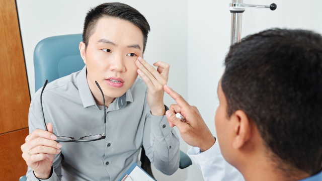
The consultation for this region, as the clinic describes it, begins by establishing which structure is responsible: orbital fat pushing forward, reduced skin elasticity, lost volume, the position of the eyelid itself, or the consequences of a previous operation. Only after that does a method come into the conversation.
An examination can do several things a discussion cannot. It can test the tone of the lower lid — StatPearls describes pulling the lid away from the eye and measuring how far it travels, releasing it to see whether it snaps back promptly, and watching whether it stays against the eye on upward gaze and with the mouth open. It can measure the position of the lid margin relative to the cornea. It can assess the tear film and note dryness, and record whether the eyelid is already sitting lower than it should. It can assess the cheek's position relative to the eye, because the midface supports the lower lid from below.
It can also find things you did not come in about. StatPearls states that every patient should have a vision examination before eyelid surgery, and notes that a history of CPAP use for sleep apnea predisposes to swelling in the cheek area, which in turn predicts more post-operative fluid there. The American Society of Plastic Surgeons, setting out what an eyelid surgery consultation covers for a United States audience, likewise expects you to be ready to discuss your general health, anything that has previously gone wrong with your eyes, what you currently take and what you have had done before, with photographs made and the options, likely outcomes and risks talked through.
Previous surgery deserves particular mention. Scarring, altered tissue planes and any change in eyelid position left by an earlier operation change what the examination finds and what can reasonably follow from it.
Why a Photograph Cannot Settle It
People increasingly try to resolve this by image — a selfie, a comparison with someone else's result, a screenshot sent for an opinion. The honest answer is that an image cannot carry the information the decision needs.
Photographs flatten. Depth in this region is read from shading, and shading is produced by the light in the room. The same under-eye can look hollow in overhead light and almost flat in diffuse light, without anything about it having changed.
The appearance is also genuinely variable. StatPearls records that lower-eyelid fullness is worse in the first hour or so after waking, when a person is tired, and after salty meals — and that with the patient lying down the fat falls back, which is exactly why patients are marked while sitting upright. An assessment has to catch the area in the posture and state it actually lives in.
And a photograph cannot be palpated, distracted, released or asked to look up. The tone of the lid, the mobility of the canthal tendons, the health of the ocular surface and the support offered by the cheek are physical findings. They are not visible in a picture at any resolution.
This is the practical reason the clinic treats the choice as diagnostic. Not to be gatekeeping about it, but because the information required is the kind you get by examining an eye.
Bringing This to a Consultation
You do not need to arrive with a conclusion. You need to arrive with a clear description of what bothers you, when you notice it most, what you have already tried, your medical and ocular history, and details of any previous eyelid surgery.
The American Academy of Dermatology suggests asking, before any cosmetic procedure, who will perform it, what their qualifications are, and what result is realistic in your particular case. For this region, three more are worth adding: which structure has been identified as responsible for what I am seeing, what would happen if the plan addressed the wrong one, and what in my examination would make a given approach unsuitable.
TAE Plastic Surgery Clinic sees patients in the Gunam Building in Haeundae-gu, Busan, and receives questions through its enquiry and appointment page. Details of the practice itself are published separately on its about the practice page.
The Bottom Line
- The clinic states that the periorbital area has thin skin with a dense concentration of blood vessels and nerves, which is why eye shape, fat quantity and skin thickness are assessed together.
- How much fat is present and where that fat sits are separate findings, and a hollow may be a true loss of volume or the lower edge of a forward bulge.
- Skin thickness and elasticity govern how tissue redrapes after anything beneath it is altered, and both change with age and with cumulative sun exposure.
- The same visible complaint commonly arises from different structures at different ages, so age informs the likeliest explanation rather than dictating a procedure.
- Lid tone, lid position, tear film and cheek support are physical findings, which is why a photograph cannot substitute for an examination.
FAQ
Why does skin thickness matter so much around the eye?
Because eyelid skin is thin, it transmits color and contour from underneath, so small structural differences are visible at the surface. Thickness and elasticity also determine how the skin settles once volume beneath it has been altered. MedlinePlus records that skin becomes thinner and less elastic with age, which is part of why the same approach does not suit every patient.
Is a hollow under my eye always lost volume?
Not necessarily. A groove can represent genuine volume loss, or it can be the boundary beneath a fat pad that is pushing forward, which reads as a hollow by contrast. The two findings have different implications, and distinguishing them is one of the things the examination is for.
Does my age decide which procedure I need?
No, though it changes what is statistically likely. Prominence of the lower-eyelid fat pads can appear in younger people with no laxity or excess skin at all, while in later decades laxity, thinning and midface change more often dominate. What matters is what your own examination finds.
Can I be assessed properly from photographs?
Photographs are useful context but cannot settle the question. Lighting alters apparent depth, lower-eyelid fullness varies with time of day, tiredness and salt intake, and the fat falls back when you lie down, which is why patients are marked sitting upright. Lid tone and lid position can only be tested in person.
What does an under-eye examination actually involve?
Assessment of eye shape, fat quantity and position, and skin thickness and quality, along with the position and tone of the lower lid, the ocular surface, and the support the cheek provides from below. StatPearls also states that every patient should have a vision examination before eyelid surgery. Previous eyelid surgery is reviewed separately, because it changes what the tissue will do.
Does a previous eyelid operation change the assessment?
Yes. Earlier surgery can leave scarring, altered tissue planes and changes in eyelid position that affect both what is found and what can sensibly follow. Bring dates, techniques where you know them, and any operative records or earlier photographs you have.
This article is for general informational purposes and does not constitute individualized medical advice, and it is not a self-diagnosis tool. Candidacy, technique, and expected outcomes can only be determined through a consultation with a qualified plastic surgeon.
References
- StatPearls: Transconjunctival Blepharoplasty
- ASPS: Eyelid Surgery Consultation
- MedlinePlus: Skin Aging
- American Academy of Dermatology: Many Ways to Firm Sagging Skin
- American Academy of Dermatology: Questions to Ask
- National Cancer Institute: Sunlight and Cancer Risk