Personalized plastic surgery, laser & anti-aging care in Haeundae, Busan
en-US

Transconjunctival Eye Surgery Korea: No Visible Scar
Transconjunctival eye surgery in Korea works through the inner eyelid. How TAE Plastic Surgery Clinic removes and grafts under-eye fat in one session.

Transconjunctival Under-Eye Surgery: Fat Removal and Grafting Through the Inner Eyelid

Medically reviewed by Dr. Kim Ki-Tae, Board-Certified Specialist in Plastic Surgery, issued by the Korean Society of Plastic and Reconstructive Surgeons
Transconjunctival eye surgery in Korea reaches the lower eyelid from behind, through the moist inner lining, so nothing is cut on the skin's outer surface. At TAE Plastic Surgery Clinic in Haeundae, Busan, the approach is used for Under-Eye Fat Correction, where protruding fat is reduced and fat harvested from elsewhere is grafted back in during the same session. This article explains how the route works, why the clinic pairs removal with grafting, and what that means for the way the area settles.
Where the incision actually goes
The conjunctiva is the thin membrane lining the inside of the eyelid. A transconjunctival approach makes its incision there, on the inner conjunctival surface, and works forward to the orbital fat from behind. The clinic describes this as carrying comparatively less burden of a visible surface scar than the alternative that cuts the skin below the lash line.
StatPearls, the peer-reviewed clinical reference published on the National Center for Biotechnology Information's Bookshelf, describes the defining feature of the technique in the same terms: the deeper structures — the orbital fat, the septum and the supporting ligaments — are reached without any skin incision at all. It also sets out the anatomy the surgeon is working within. Below the tarsal plate, the lower eyelid is effectively three layers: an outer layer of skin and the orbicularis muscle, a middle layer formed by the orbital septum and the eyelid retractors, and an inner layer of conjunctiva. The orbital fat sits between the septum and the retractors, and it arrives in three distinct pads — medial, central and lateral — separated by the inferior oblique muscle and by a thickening in the septum.
That detail is not trivia. It explains why the operation is described as reducing bulges rather than removing a single lump, and why the lateral pad in particular has to be deliberately exposed rather than assumed to be reachable.
Why removal and grafting happen together
The clinic's stated point of difference is that it removes the protruding fat and then grafts fat back, rather than sliding existing fat downward. Fat harvested from a donor area is processed before transfer, with cells taken from the superficial layer close to the skin surface used specifically because the tissue around the eye is thin. The clinic states that this can help produce a comparatively natural contour in an area where thicker material would read as a ridge.
The logic is anatomical. Many people who dislike their under-eye area have two things happening at once: a pad pushing forward, and a groove sitting back beneath it. Address only the forward part and the groove can become more conspicuous, because the bulge above it was providing a visual transition. Restoring volume where volume is missing is what keeps the corrected area continuous with the cheek rather than stepping down into it.
The equipment reflects that. The clinic describes a laser-assisted transconjunctival approach working through a micro-incision on the inner eyelid, and characterizes the procedure as a skin-incision-free technique intended to reduce scarring and recovery time.
Who the approach suits, and who it does not
The clinic considers this procedure for people whose under-eye shows orbital fat protruding forward, or the combined pattern where the area looks bulging and hollow simultaneously, and it assesses eye shape, the volume of fat and skin thickness together. The reason given is structural: the tissue around the eye is thin and densely supplied with blood vessels and nerves.
The clinical literature adds a set of limits that matter here. StatPearls lists skin excess, poor lower eyelid skin tone, redundancy of the orbicularis muscle, festoons, lower eyelid laxity, existing ectropion, eyelid retraction and significant cheek descent among the situations where a purely conjunctival procedure on its own is not the appropriate answer, and notes that the technique in its pure form suits isolated prominence of the fat pads. It describes a series of clinical tests — pulling the eyelid away from the globe and watching how readily it returns, for instance — used to establish whether the lid has enough tone to tolerate the procedure without support.
The clinic's own exclusions sit alongside those. Pregnancy and breastfeeding rule the procedure out for the time being. So do medical conditions that are not under control and could interfere with the operation or with healing, and any active infection in the area around the eye. Expectations that the anatomy cannot deliver are treated the same way. Whether to proceed is decided by the doctor once your health, history and goals have been assessed individually.
The alternative route, and the reason it is weighed carefully
The comparison most patients are implicitly making is against the version of lower eyelid surgery that incises the skin outside the eye. The clinic's own material notes that this external approach may carry a burden regarding scarring, and that consideration is also needed for the possibility of ectropion — the eyelid turning outward — during the process of pulling and fixing the fat, as well as for fat protruding again over time.
Ectropion is the complication US patients most often ask about by name, and it is worth understanding rather than simply fearing. The clinical reference describes it as one of the malpositions that can follow when the lower eyelid's support is disturbed: scarring or a shortage of skin in the outer layer can pull the lid away from the eye, and the same result can follow from weakness in the muscle or from stretching of the tendons that anchor the lid at its inner and outer corners. The lid normally sits snugly against the globe, at or just above the lower edge of the iris, and it does so because several structures are intact at once.
None of this makes the external approach wrong; it has its own indications, particularly where skin excess is the dominant problem. It does explain why the route is chosen against the examination rather than chosen first.
What the surgery and the days afterward involve
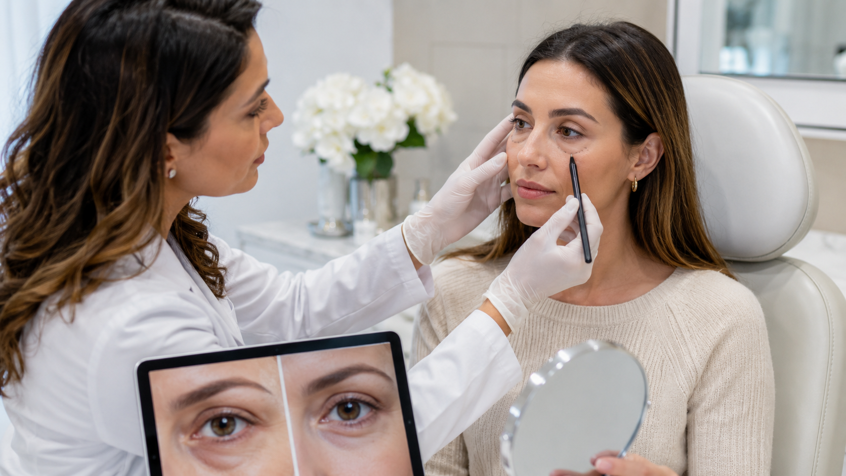
The sequence the clinic describes runs as follows: assessment of the under-eye structure and the balance between protrusion and hollowing; planning how much fat to remove and how much to graft back; anesthesia; the incision on the inner conjunctival surface; removal of the protruding orbital fat; harvest and processing of donor fat; grafting into the under-eye to restore volume; and recovery-room observation with discharge instructions. Anesthesia is typically local with sedation, or general depending on the extent of surgery. The procedure itself typically takes one to three hours depending on complexity.
The opening few days carry the heaviest bruising and puffiness, which then eases across the fortnight that follows; light routine activity is usually manageable again somewhere around day seven to day ten. How quickly any given person gets there depends on skin type, tissue characteristics and general health, and it is checked at follow-up rather than assumed. Genuine change becomes readable once the bulk of the swelling has gone, at roughly two to three weeks, while the settled outcome generally arrives near the three-month mark and is described as long-lasting to permanent.
Getting ready means pausing blood-thinning drugs and supplements a week or two ahead, setting cigarettes and alcohol aside, fasting as instructed before anesthesia, and having routine pre-operative bloods done. Afterward the instructions run: keep the site clean and shielded, hold off on hard exercise, heavy lifting, drinking and smoking for somewhere between two and four weeks, sleep propped up where that applies, and attend every scheduled check. Written guidance setting out the day-by-day timeline goes to each patient.
MedlinePlus, the patient-education service of the U.S. National Library of Medicine, sets out general expectations for the period after any operation, including the value of following wound-care instructions precisely and of knowing which symptoms warrant contacting the surgical team rather than waiting. That framing is useful for anyone recovering at a distance from where they were treated.
Risks specific to this route
The clinic's list for this procedure begins with the expected: short-lived swelling and bruising, plus discomfort. Beyond that it names bleeding and infection, unevenness between the two sides, irregularities in the contour, fat that remains prominent or a hollow that persists or returns, correction that goes too far or not far enough, and altered sensation. A small number of outcomes sit in a more serious category and occur rarely: the eyelid coming to sit in the wrong position, dry eye that does not resolve, disturbance of vision, and damage to the structures surrounding the eye socket.
The clinical literature's own complication list for the conjunctival approach overlaps and adds detail: chemosis, which is swelling of the conjunctiva itself; infection; bleeding; residual prominence of the fat pads; lower eyelid retraction; excessive fat removal; temporary nerve irritation; injury to the extraocular muscles, most commonly the inferior oblique; double vision; and, very rarely, loss of vision. It also notes that conservative fat reduction is preferred precisely because removing too much is difficult to undo.
Outcomes cannot be guaranteed. What can reasonably be discussed in advance is which pattern your anatomy shows, which structures are being worked on, and what the realistic ceiling is for your starting point.
Preparing for the consultation
The American Academy of Dermatology recommends establishing, before any cosmetic procedure, who will carry it out, what training they hold, and what result is realistic for your individual case. For under-eye work it is also worth asking how much fat is planned for removal against how much for grafting, whether your lid tone was formally tested, and what the plan would be if the result settles unevenly. A clinic overview covers how the practice is organized. Questions about your own eyelids go through the enquiry form.
The Bottom Line
- Transconjunctival under-eye surgery reaches the orbital fat through an incision on the inner lining of the eyelid, so no cut is made on the skin's outer surface.
- TAE Plastic Surgery Clinic pairs fat removal with grafting in the same session, using finely processed fat from the superficial layer because the tissue around the eye is thin.
- A purely conjunctival approach suits isolated prominence of the fat pads; skin excess, eyelid laxity, festoons or existing retraction point toward a different plan.
- Swelling settles over one to two weeks, visible change appears at two to three weeks, and the settled result is typically seen at around three months.
FAQ

Where exactly is the incision made?
On the inner conjunctival surface of the lower eyelid — the moist lining on the inside — so the surgeon reaches the orbital fat from behind rather than through the skin. The clinic describes this as carrying comparatively less burden of a visible surface scar.
Why is fat both removed and put back?
Because the two problems often occur together. Fat can push forward while the groove beneath it sits back, and reducing only the forward portion can make the hollow more noticeable. Grafting processed fat into the deficient area is what keeps the corrected contour continuous with the surrounding face.
Why use fat from the superficial layer?
The clinic states that cells from the layer close to the skin surface are finely processed before transfer specifically because the tissue around the eye is thin, and that this can help produce a comparatively natural contour in that setting.
What anesthesia is used?
Typically local anesthesia with sedation, or general anesthesia depending on the extent of the surgery. The exact approach is confirmed with you at consultation based on your individual comfort and medical history.
Am I a candidate if I have loose lower eyelid skin?
Possibly not for this approach on its own. The clinical literature lists skin excess, poor lower eyelid tone, orbicularis redundancy, festoons and lower eyelid laxity among the findings that make a purely conjunctival procedure inappropriate by itself, which is why lid tone is examined before a method is settled.
What are the serious risks?
Beyond the expected swelling and bruising, possibilities include bleeding, infection, asymmetry, contour irregularity, over- or under-correction, sensory change, and rarely eyelid malposition, persistent dry eye, visual disturbance or injury to surrounding orbital structures. The clinical literature adds chemosis, lower eyelid retraction, injury to the inferior oblique muscle and double vision.
How long until I can go back to normal activity?
Most people return to light daily activity within a week to ten days, with strenuous exercise, heavy lifting, alcohol and smoking avoided for two to four weeks. The clinic confirms the individual schedule at consultation.
This article is for general informational purposes and does not constitute individualized medical advice, and it is not a self-diagnosis tool. Care described here is delivered in the Republic of Korea under Korean medical licensure rather than United States licensure, and the clinic is closed on Sundays and observes a daily lunch break, which affects scheduling for short international trips. Speak with a qualified plastic surgeon about your own circumstances before making any treatment decision.
References
- StatPearls: Transconjunctival Blepharoplasty
- MedlinePlus: After Surgery
- MedlinePlus: Plastic and Cosmetic Surgery
- MedlinePlus: Wounds and Injuries
- American Academy of Dermatology: Questions to Ask
- American Academy of Dermatology: Cosmetic Treatments